


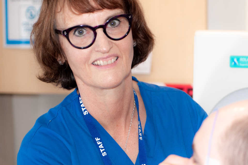
A former dispensing optician, Juliet Ware is Auckland’s only ophthalmic electrodiagnostic technician working in the public healthcare setting. She is based in Greenlane Clinical Centre’s ophthalmology department.
Serendipity put Ware on the path to visual electrodiagnostics*. “I was working crazy retail hours, managing and dispensing at a large OPSM branch. I then did some optical repping to get out of the retail environment, which required a lot of out-of-town travel.” Ready for a change, Ware learned from her neighbour, a Greenlane eye clinic nurse, that they had an opening for a trainee ophthalmic electrodiagnostic technician. “I decided to apply, even though I didn’t really know what it was about. Here I am, over 10 years later!”
Retraining on the job had its challenges, electrodiagnostics being completely different to optics, she said. “I was very lucky as I trained under Dr Dianne Sharp, although she must have torn her hair out in despair. Dr Sharp is one smart cookie, but she really explained stuff to me in a way I could understand.”
Ware sees three to six patients a day, with tests typically taking between 30 and 60 minutes each, with many patients requiring a combination of tests to give complete information about their visual problem, she explained. While most have either retinal or optic nerve issues, she said roughly a third of patients require toxicity monitoring as they are on hydroxychloroquine, prescribed mainly for lupus and rheumatoid arthritis.
A challenging aspect of the role is performing tests such as an electroretinogram, which require electrodes placed around the face and in the eyes. This can be extremely difficult with young children and babies as they don’t understand what’s going on, Ware said. “Sometimes, as a last resort with an uncooperative child, we end up performing the test in theatre under general anaesthetic.”
Being the only ophthalmic electrodiagnostic technician in the Auckland region can also add pressure, Ware admitted. “I get really stressed if I get sick or have to take unplanned leave as there is no one able to step in and perform the full range of tests. I feel especially bad if a patient is coming from out of town and it’s too late to stop them.” To solve this conundrum and to help cut the waitlist, Ware is currently training one of Greenlane’s orthoptists in her duties.
Aside from adhering to the International Society for Clinical Electrophysiology of Vision’s test standards, being approachable, relatable and flexible are key to the job, according to Ware. “I must make sure the patient is comfortable, at ease and given clear instructions so that they can perform the tests to the greatest of their ability.”
Patients are often very grateful to get the procedure done, inching one step closer to a diagnosis, she added. “I see patients from all walks of life and ages and love working with them. I find the interaction very rewarding.”
If she could change one thing about her work at Greenlane, she joked it would be to get Dr Sharp back! “In all seriousness, I do miss her expertise, but consider myself lucky to have other amazing doctors I work with, including Drs Leo Sheck and Tracey Wong.”
When not busy at the clinic, Ware said she loves running, having completed six full marathons and numerous half marathons. “The Auckland and Rotorua full marathons are my most memorable races. The Rotorua one is especially stunning; you run around Lake Rotorua and the views are just incredible.” A devoted animal-rights advocate, she also keeps busy fostering animals, with a Jack Russell, one cat, eight rabbits and one foster bunny currently under her roof.
*Visual electrophysiology is a range of techniques used to test the functioning of cells along the full visual pathway – from the retina to the optic nerve to the brain’s primary visual cortex. It measures and records electrical signals in these areas in response to visual stimuli. Results are used to help detect, diagnose and evaluate visual disorders including retinitis pigmentosa, inherited-retinal degeneration and macular degeneration.