


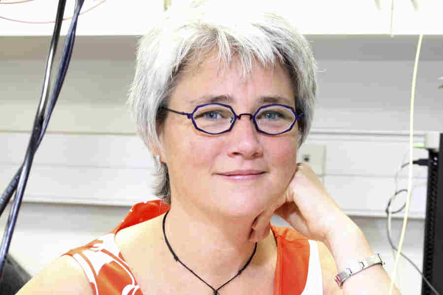
It can be easy to get stuck in a professional silo, but research lead by physicist Dr Frederique Vanholsbeeck is a reminder that no man – or specialism - is an island. Her team, based in the University of Auckland’s physics department, are working on the development of new differentiation tools in optical coherence tomography (OCT).
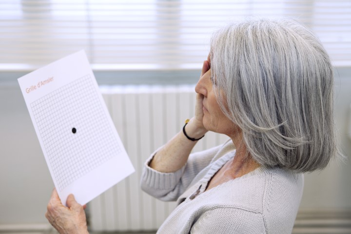
“OCT is a versatile and unique technique with many applications,” explains Dr Vanholsbeeck. “But it lacks discrimination in key areas. We are monitoring chromatic dispersion and tissue stiffness and looking at how new analysis and measuring techniques can offer more accurate results to differentiate tissue or detect early signs of diseases.”
Chromatic dispersion blurs the contours in OCT images, making it hard to gain accurate measurements of choroidal thickness. But by combining sophisticated data analysis techniques with a new optical laser, Dr Vanholsbeeck is aiming to enhance the accuracy, and therefore the value of OCT.
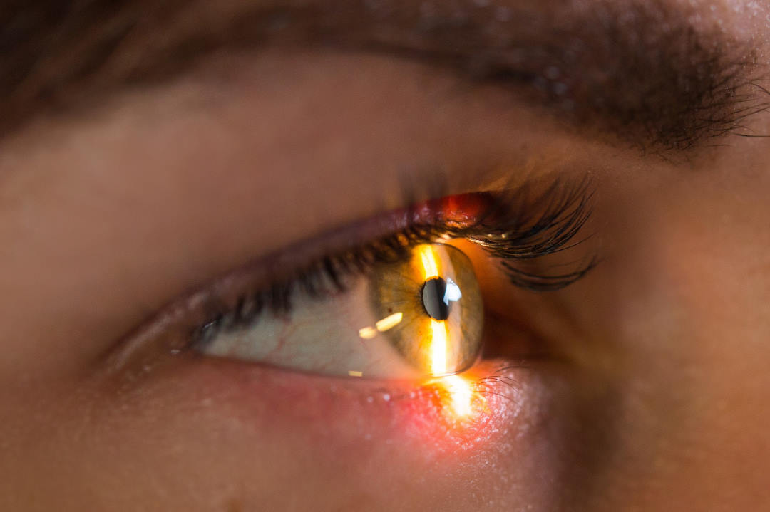
“If you analyse the light after it has interacted with tissue, you can extract information about your sample such as its composition,” says the Brussels-born physicist who studied nonlinear optics as part of her PhD before going into biomedical imaging and biophotonics. “We have a Marsden Research grant [awarded November 2015] and a team of five, including an optometrist, a post-doc and two students as well as overseas collaborators. We already have results and will be publishing our first paper soon.”
The potential of this research goes beyond simply improving OCT imaging systems. The results will enable clinicians to look deep into their patient’s eye, leading to potential new therapeutic applications globally.
If you’re a student interested in working with Dr Vanholsbeeck, contact her by email; f.vanholsbeeck@auckland.ac.nz or check out more about her research group at www.biophotonics-newzealand.com