


The 2007 Tear Film & Ocular Surface Society Dry Eye Workshop (TFOS DEWS)1 marked a major milestone in standardising the understanding and management of dry eye disease (DED). A decade later, TFOS DEWS II (2017)2 expanded on this foundation, supported by widespread global research. Now, TFOS DEWS III represents the next step in this evolution, reflecting the significant scientific advancements since DEWS II, in refining the definition, classification, diagnosis and management of DED.
Definition
TFOS DEWS III redefines DED as: a multifactorial, symptomatic disease characterised by a loss of homeostasis of the tear film and/or ocular surface, in which tear film instability and hyperosmolarity, ocular surface inflammation and damage and neurosensory abnormalities are aetiological factors.
This updated definition acknowledges that tear film and ocular surface homeostasis may be disrupted by various pathophysiological mechanisms, any of which can be present in an individual.
Diagnostic methodology
Diagnosing DED has long been a clinical challenge. Differential diagnosis is essential to rule out conditions that mimic DED, and a combination of both symptoms and signs should be used to confirm a diagnosis.
Symptoms
To standardise symptom assessment, TFOS DEWS III recommends use of a single, validated screening tool. While multiple questionnaires have previously been used, a shortened form of the Ocular Surface Disease Index (OSDI), the OSDI-63 has been selected for its sensitivity, simplicity and practicality in routine care. A score of ≥4 indicates further diagnostic testing is warranted4. See https://heiko-pult.de/media/files/OSDI-6-EN-NEW.pdf
Signs

Fig 1. TFOS DEWS III Diagnostic criteria for dry eye
If symptoms are present, objective markers of tear film homeostasis support a positive diagnosis. An abnormal result in any one of the following three tests confirms DED (Fig 1):
Contrasting with TFOS DEWS II – which required all three of these tests to be confident a patient did not have DED – TFOS DEWS III allows for the absence of DED to be confirmed by performing only two clinical tests, provided one of them is comprehensive ocular surface staining (that includes corneal, conjunctival and lid margin staining). Additionally, where non-invasive testing is unavailable, fluorescein tear film breakup time may be used as a proxy, the diagnostic cut-off reduced from 10 to 5 seconds due to better understanding of fluorescein’s impact on tear film stability4.
TFOS DEWS III Digest
The TFOS DEWS III Digest summarises interdisciplinary research across several key areas of DED including epidemiology, pathophysiology, pain and sensation, and sex, gender and hormones. These inform both diagnostic and therapeutic frameworks.
Epidemiology
DED prevalence varies significantly across populations, reaching up to 62.9% in certain populations5. DEWS III classifies risk factors as conclusive or probable risks for DED, categorising them as either modifiable or non-modifiable. Broad categories include demographics, lifestyle/environment, systemic disorders, ocular disorders, medications and iatrogenic factors5. Identifying these factors is crucial in both preventing and managing DED.
Pathophysiology
TFOS DEWS III solidifies distinctions between aqueous deficient and evaporative dry eye. While inflammation is a recognised driver, evaporative dry eye often presents with minimal or no inflammatory markers present in tears6. Inflammation should therefore not be assumed to be present in all DED cases, although it may emerge secondarily from stress on the ocualr surface induced by triggering the vicious circle of dry eye. Immune-mediated aetiologies eg. Sjögren’s disease, Stevens-Johnson syndrome and ocular graft versus host disease (GVHD) are associated with more severe disease presentation7, where inflammation is a primary mechanism.
Pain and sensation
The ocular surface is densely innervated with sensory nerves. The TFOS DEWS II pain and sensation report8 outlined how trauma, surgery, systemic disease and aging can disrupt corneal nerve architecture, impairing protective reflexes such as blinking and tearing. TFOS DEWS III explores recent findings in corneal nerve modelling and the relevance of the anatomical and functional status of corneal nerves in diagnosing and managing DED5.
Sex, gender and hormones
Sex- and gender-related differences influence susceptibility to DED, as highlighted in TFOS DEWS II9 and expanded upon in DEWS III5.These differences are attributed to hormones, sex chromosomes, sex-specific autosomal factors, epigenetics, care-seeking behaviours and service utilisation. New findings reveal that androgens, progesterone, thyroid eye disease and insulin/insulin-like growth factor pathways can significantly affect lacrimal gland, meibomian gland, corneal and eyelid function.
Subclassification

Fig 2. After dry eye diagnosis, nine subtypes representing tear film deficiencies, eyelid anomalies and ocular surface abnormalities, are checked and managed as required, to restore tear film and ocular surface homeostasis
DED commonly arises as a downstream consequence of a range of aetiologies. TFOS DEWS III classifies DED based on aetiological subtypes, aiding targeted management4. Broadly, these include tear film component deficiencies, eyelid anomalies, ocular surface abnormalities and systemic conditions. Assessment techniques for each subtype of DED are outlined in the TFOS DEWS III Diagnostic Methodology report.
Tear film component deficiencies
The tear film consists of lipid, aqueous and mucin components. Deficiencies in lipid or aqueous lead to the well-established evaporative and aqueous deficient dry eye subtypes, respectively. Mucins play a critical role in tear film spread and ocular surface protection10 and likely arise from secondary ocular surface inflammation leading to cellular damage11.
Eyelid anomalies
Blinking plays a critical role in tear distribution and debris removal4. Abnormal blinking patterns due to lifestyle factors or conditions influencing eyelid function have been implicated in DED development and exacerbation12,13. Diseases of the eyelid, such as anterior blepharitis and meibomian gland dysfunction (MGD), remain widely recognised as common drivers of DED through microbial overgrowth and impaired lipid secretion, respectively4.
Ocular surface abnormalities
Ocular surface abnormalities include anatomical misalignment (eg. due to pterygium, pinguecula and conjunctivochalasis), abnormal corneal sensitivity and cellular damage, which can hinder tear film production, spread and blinking, leading to a loss of tear film and/or ocular surface homeostasis4.
Systemic diseases
Systemic conditions (eg. autoimmune, inflammatory, metabolic and neural) can significantly impact ocular surface health through inflammation, metabolic dysregulation and ocular surface exposure. This underscores the need for interdisciplinary collaboration where systemic disease drives DED4.
Management and therapy
DED management is rapidly evolving; the TFOS DEWS III Management and Therapy14 report outlines evidence-based strategies in three concise tables. Management spans low-risk, at-home interventions through to more invasive medical or surgical options, aligned with individual aetiologies.
Lifestyle modifications remain paramount across all dry eye subtypes, as outlined in the TFOS Lifestyle reports15, where they influence not only ocular health, but overall wellbeing and quality of life. Their consideration remains critical in both the development and management of DED.
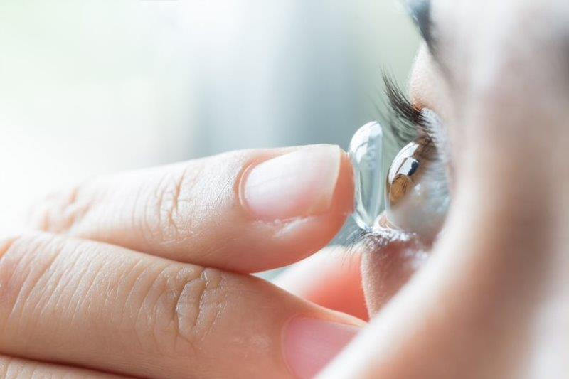
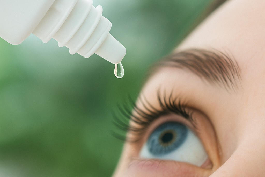
Tear supplementation and stabilisation remain the mainstays of therapy, with growing options to restore, stimulate or stabilise tear film components.
TFOS DEWS III discussed the use of artificial tears, where more novel ingredients, such as trehalose and perfluorohexyloctane, offer broader options for tear film stabilisation and ocular surface protection, healing and lubrication. Neurostimulation/neuromodulation targeting the sensory nerves has also been demonstrated to increase tear production6,16, potentially providing an alternative to artificial tears. Blinking therapy may help to improve spontaneous blinking behaviour and promote lipid secretion and tear film spread17.
A range of pharmacological and naturally derived agents, including anti-inflammatories, blood-derived products and amniotic membrane products, show promise in managing more severe DED.
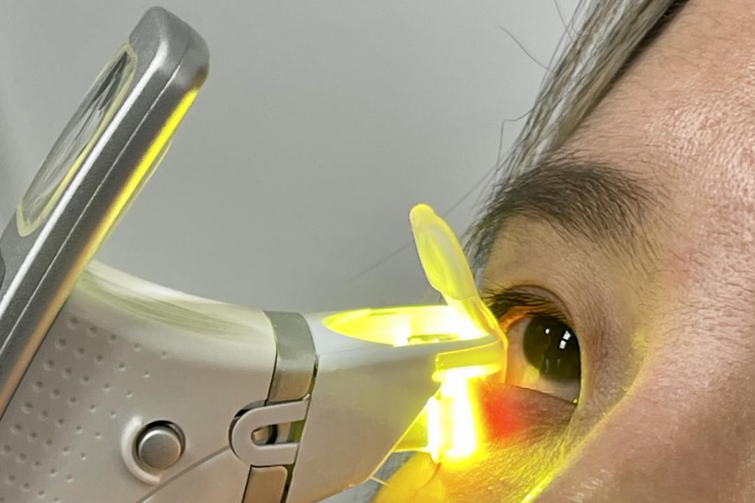
MGD is recognised as a common driver of DED, with many treatments designed to specifically target the eyelids and meibomian glands. Various heat-based, light-based and electrotherapeutic devices increase treatment options available to clinicians14.
In more severe DED that is non-responsive to other management approaches, surgical options may help to address anatomical misalignment, eyelid abnormalities and severe aqueous deficient dry eye14.
Summary
TFOS DEWS III represents a significant evidence-based resource and advance in DED clinical care recommendations. By updating definitions, refining diagnostic criteria, describing advances in our understanding of pathophysiological mechanisms and emphasising personalised, evidence-based management, it sets a new benchmark for improving patient outcomes and reducing the global burden of DED.
References
1. Lemp MA, Baudouin C, Baum J, Dogru M, Foulks GN, Kinoshita S, et al. The definition and classification of dry eye disease: Report of the Definition and Classification Subcommittee of the international Dry Eye WorkShop (2007). Ocular Surface. 2007;5:75-92.
2. Craig JP, Nelson JD, Azar DT, Belmonte C, Bron AJ, Chauhan SK, et al. TFOS DEWS II Report Executive Summary. Ocul Surf. 2017;15:802-12.
3. Pult H, Wolffsohn JS. The development and evaluation of the new Ocular Surface Disease Index-6. The ocular surface. 2019;17:817-21.
4. Wolffsohn JS, Benítez-Del-Castillo J, Loya-Garcia D, Inomata T, Iyar G, Liang L, et al. TFOS DEWS III Diagnostic Methodology. American journal of ophthalmology. 2025.
5. Stapleton F, Argüeso P, Asbell P, Azar D, Bosworth C, Chen W, et al. TFOS DEWS III Digest Report. American journal of ophthalmology. 2025.
6. Perumal N, Funke S, Pfeiffer N, Grus FH. Proteomics analysis of human tears from aqueous-deficient and evaporative dry eye patients. Scientific reports. 2016;6:29629.
7. Chotikavanich S, de Paiva CS, Chen JJ, Bian F, Farley WJ, Pflugfelder SC. Production and activity of matrix metalloproteinase-9 on the ocular surface increase in dysfunctional tear syndrome. Investigative ophthalmology & visual science. 2009;50:3203-9.
8. Belmonte C, Nichols JJ, Cox SM, Brock JA, Begley CG, Bereiter DA, et al. TFOS DEWS II pain and sensation report. The ocular surface. 2017;15:404-37.
9. Sullivan DA, Rocha EM, Aragona P, Clayton JA, Ding J, Golebiowski B, et al. TFOS DEWS II Sex, Gender, and Hormones Report. Ocul Surf. 2017;15:284-333.
10. Hori Y. Secreted mucins on the ocular surface. Investigative ophthalmology & visual science. 2018;59:DES151-DES6.
11. You Y, Chen J, Chen H, Wang J, Xie H, Pi X, et al. Investigation of Conjunctival Goblet Cell and Tear MUC5AC Protein in Patients With Graves’ Ophthalmopathy. Translational vision science & technology. 2023;12:19-.
12. Su Y, Liang Q, Su G, Wang N, Baudouin C, Labbé A. Spontaneous eye blink patterns in dry eye: clinical correlations. Investigative ophthalmology & visual science. 2018;59:5149-56.
13. Mitchell T, Murri M, Pflugfelder SC. Video viewing blink rate in normal and dry eyes. Eye & contact lens. 2021;47:442-4.
14. Jones L, Craig JP, Markoulli M, Karpecki P, Akpek EK, Basu S, et al. TFOS DEWS III Management and Therapy Report. American journal of ophthalmology. 2025.
15. Craig JP, Alves M, Wolffsohn JS, Downie LE, Efron N, Galor A, et al. TFOS lifestyle report executive summary: a lifestyle epidemic-ocular surface disease. The ocular surface. 2023;30:240-53.
16. Gumus K, Pflugfelder SC. Intranasal tear neurostimulation: an emerging concept in the treatment of dry eye. International ophthalmology clinics. 2017;57:101-8.
17. Ashwini D, Ramesh SV, Nosch D, Wilmot N. Efficacy of blink software in improving the blink rate and dry eye symptoms in visual display terminal users–A single-blinded randomized control trial. Indian journal of ophthalmology. 2021;69:2643-8.

Professor Jennifer Craig is head of the Ocular Surface Laboratory in the University of Auckland’s Department of Ophthalmology, was the former chair of the TFOS Lifestyle Workshop and is now the chair (formerly called president) of TFOS DEWS II.

Jordan Cooper is a former optometry graduate and current PhD candidate with the OSL, working under the supervision of Professor Jennifer Craig.